


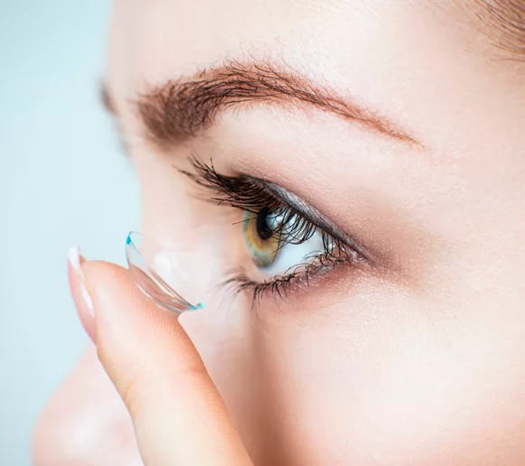
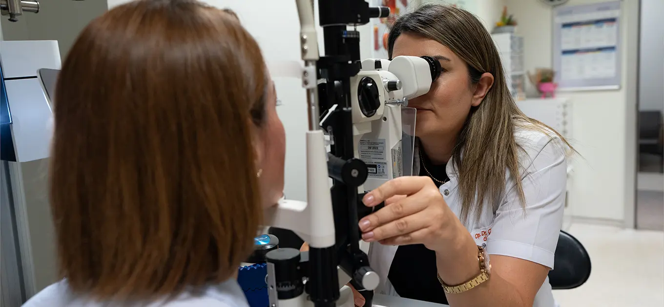
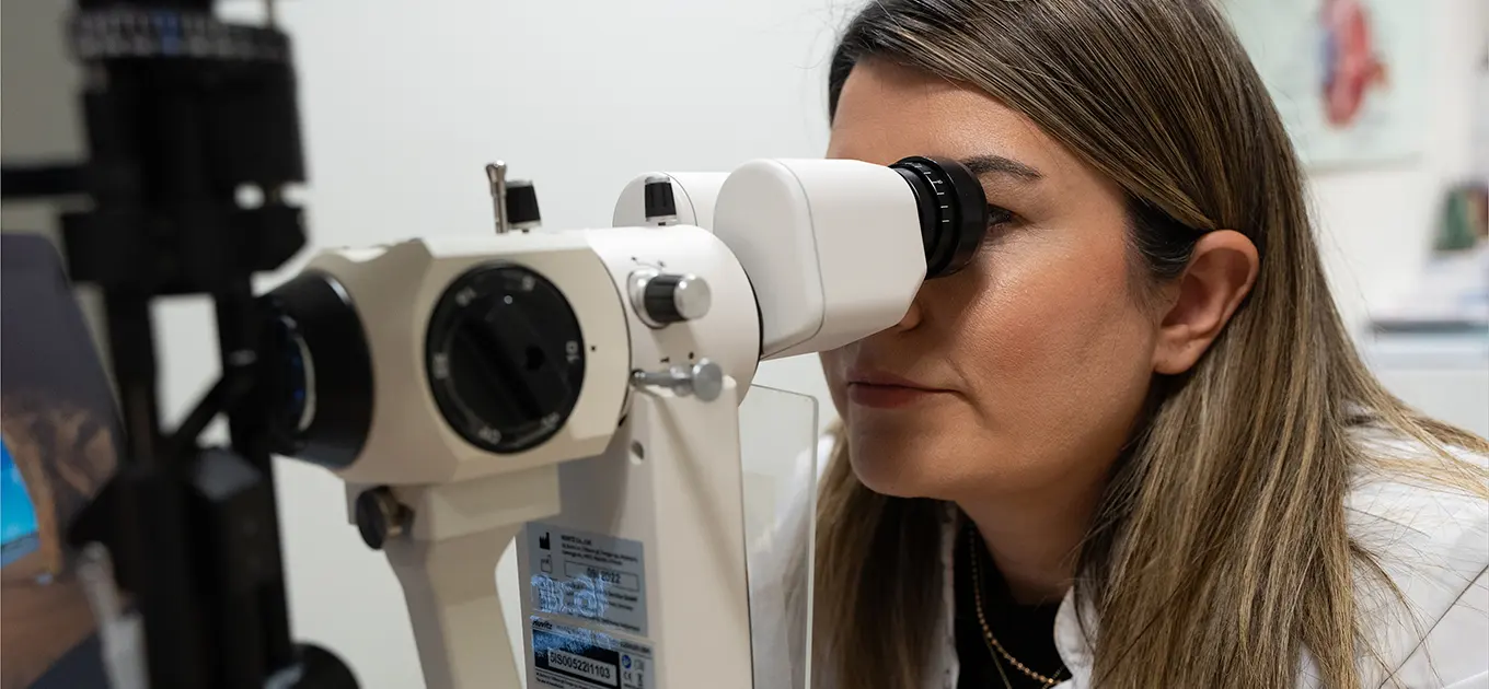
Thanks to the computerized eye health diagnostic systems we use in our Ophthalmology clinic, existing conditions in patients such as myopia, hyperopia, and astigmatism can be diagnosed, and appropriate glasses or contact lenses can be prescribed.

In patients with diabetes, poor blood sugar control can damage blood vessels and cause retinal haemorrhages. If left untreated, these haemorrhages may spread within the eye and lead to permanent vision loss. In our clinic, such conditions can be detected early through fundus examination after pupil dilation. With optical coherence tomography (OCT), oedema in the visual centre can be identified and treated with intravitreal injections. Retinal angiography also allows detection of vessels at risk of bleeding, which can be treated with retinal laser therapy.
Macular degeneration (yellow spot disease) is a leading cause of vision loss, especially in elderly patients. With the help of OCT and retinal angiography, it can be detected at an early stage, and its progression can be slowed with intravitreal injection treatments when necessary.
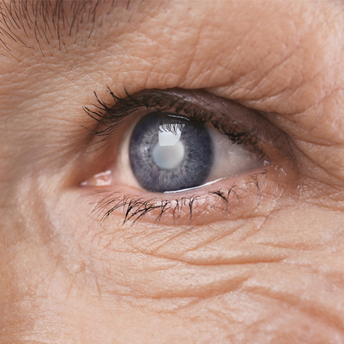
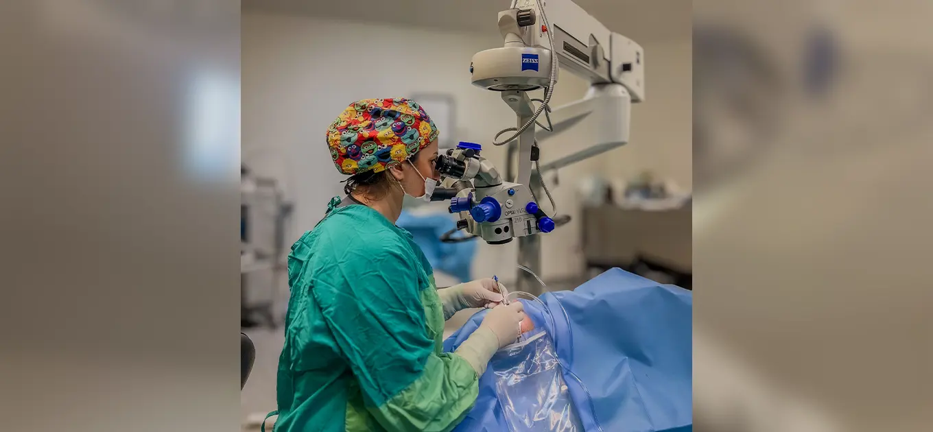
The cataractous lens is removed using the Phaco method (Phacoemulsification) and an artificial lens is placed in its position through a sutureless surgery.
With advancing age, cataract formation is common, which involves the loss of transparency of the eye's lens. Once a cataract has formed, it cannot be corrected by any means other than surgical intervention. In our Ophthalmology department, we apply the PHACO method to remove the cataractous lens, and an artificial lens is placed in the position of the removed natural lens of the eye.

Glaucoma is the second most common cause of vision impairment in the world after macular degeneration. Glaucoma is a disease that progresses insidiously without any symptoms, damaging the optic nerve and leading to permanent vision loss. The optic nerve damage caused by glaucoma cannot be reversed. Therefore, especially after the age of 40, patients with a family history of glaucoma or diabetes should have a routine comprehensive examination, even if they have no symptoms. Thanks to optical coherence tomography, optic nerve damage can be detected at a very early stage, and intraocular pressure can be controlled before the disease has progressed. Additionally, thanks to the special software of the tomography device used in our hospital, glaucoma progression can be monitored by comparing previous measurements during the patient's follow-up examinations.
Especially with aging, due to deterioration of the eyelid condition, the eyelashes may turn inward or the eyelids may turn outward from the eye. In both cases, burning, irritation, redness, and even tissue deterioration can occur in the eye, as the protective mechanism of the eyelids is compromised. In these cases, the eyelids can be restored to their previous position using special surgical techniques.
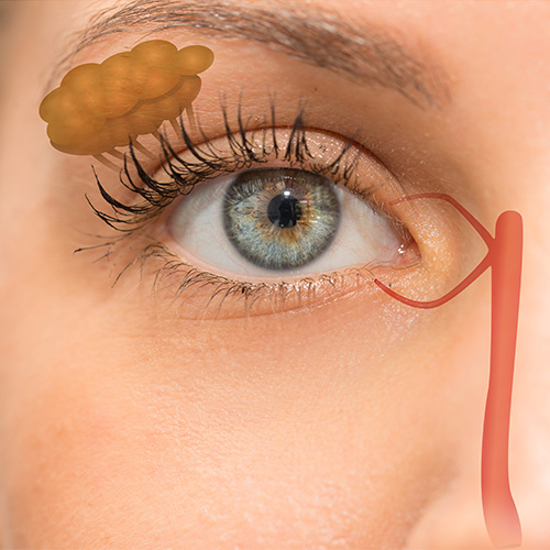
The tear duct starts from the lacrimal sac and passes toward the nose. A blockage in any part of this duct can be permanently corrected by attaching a temporary silicone tube to the area.
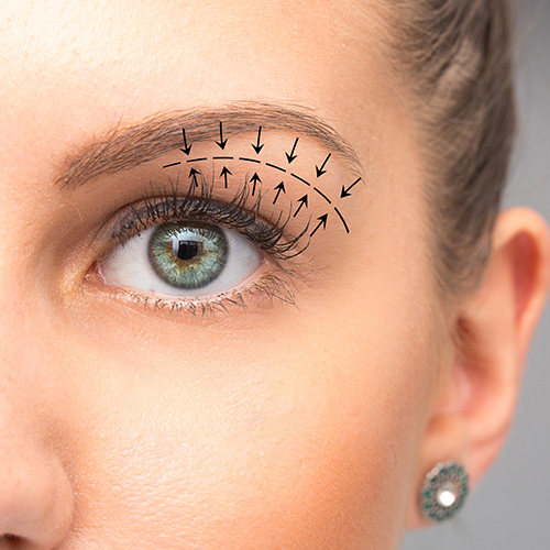
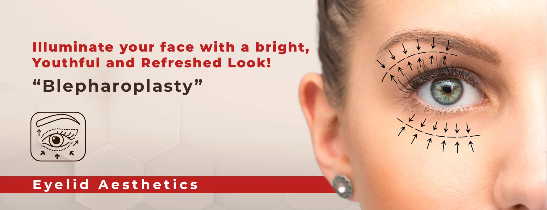
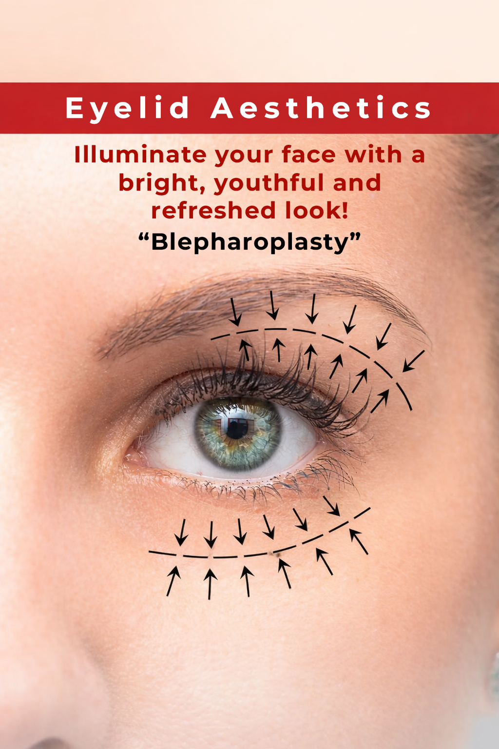
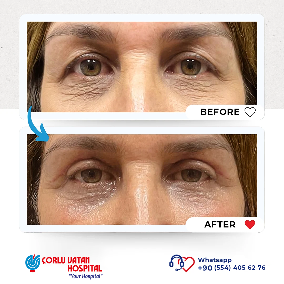
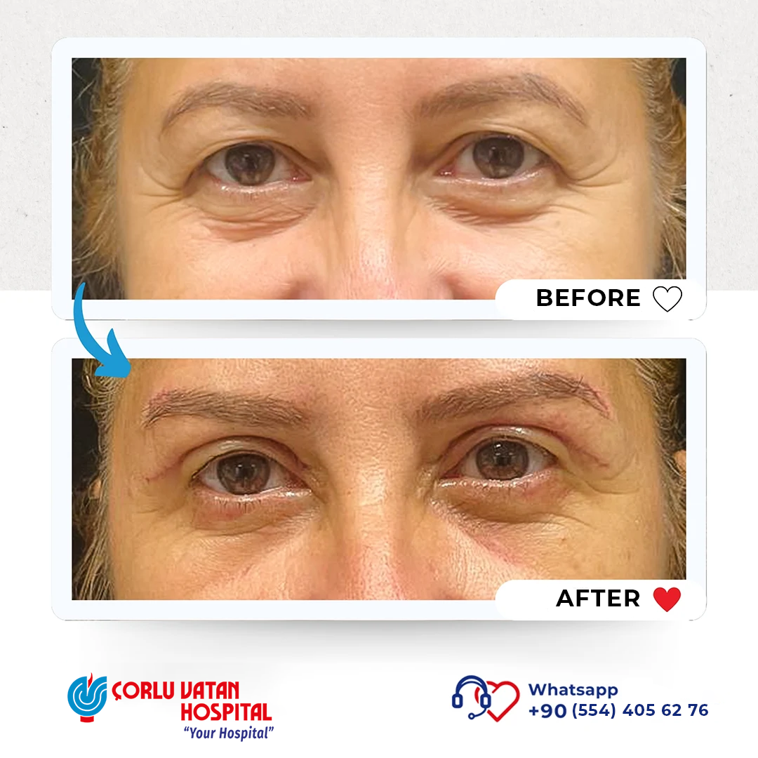
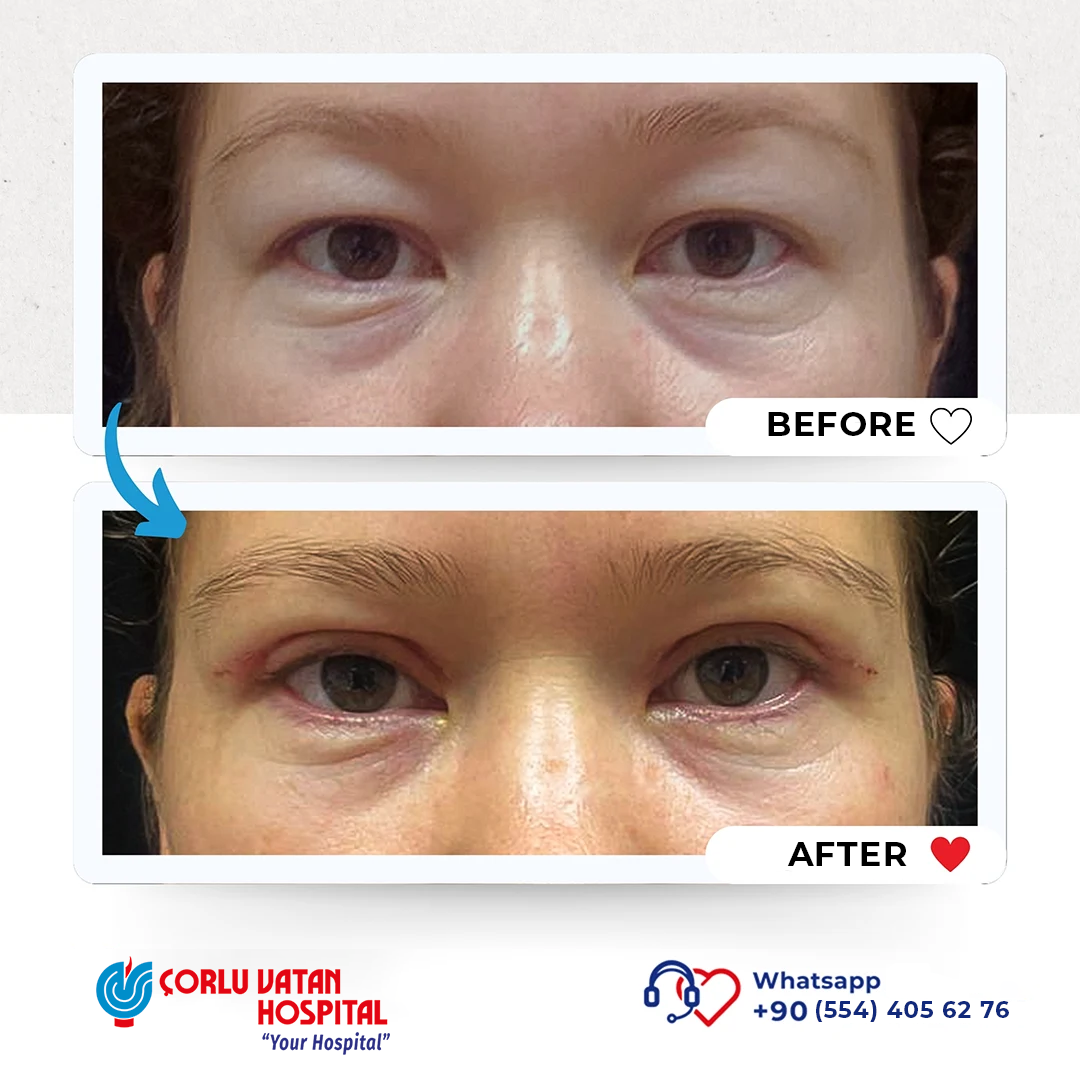
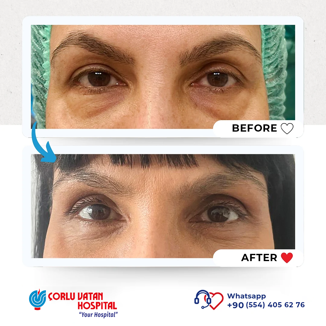
Upper eyelid blepharoplasty is a surgery performed when there is loose and excess skin and fatty deposits in the upper eyelid area, with the main cause being the aging process, and sometimes structural or pathological reasons. After a detailed physical examination and explanation, a surgical intervention plan is prepared. During the surgery, the incision line is planned to correspond to the upper eyelid crease, and during the intervention, excess muscle and fatty tissue are removed. Then, self-absorbing sutures are placed. In this surgery, scars are hidden in the eyelid crease so they are not visible. During the initial consultation with the patient, features such as drooping eyebrows and other possible problems in the periocular area are also evaluated, and options for their correction during the surgical intervention are discussed.
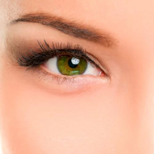
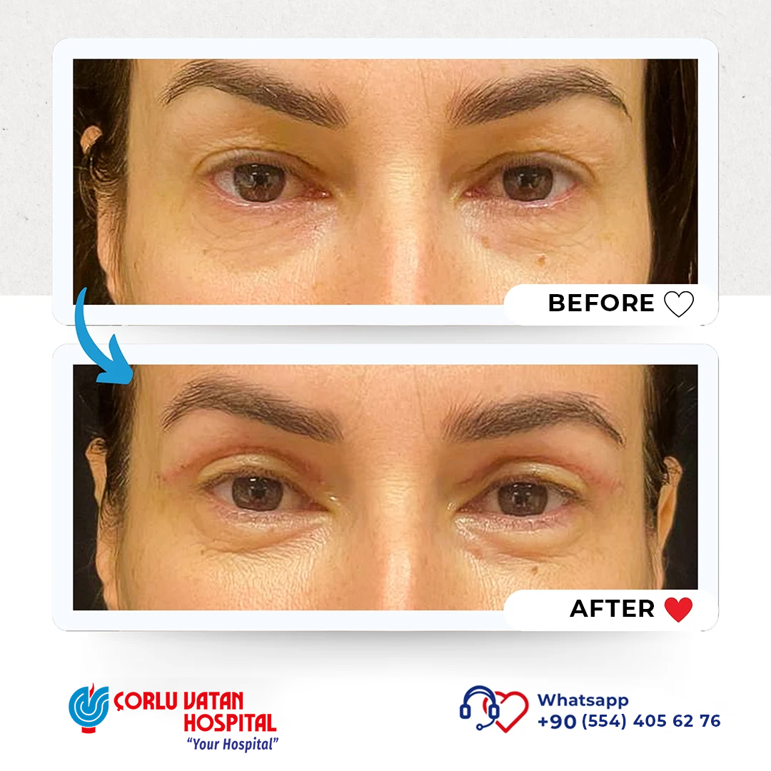
Almond eye surgery is a procedure that lifts the outer corners of the eyes to create a more youthful and refreshed appearance. By elevating the outer eyelid area, the eyes gain a more defined shape, and the tired facial expression can be reduced. After evaluating the patient’s anatomy and expectations, the most appropriate treatment plan is determined.
In our hospital, the procedure can be performed using two methods. The first is a surgical technique, where the outer corner of the eyelid is lifted and fixed to the bone membrane with permanent sutures.
The second is a non-surgical method using meso-threads, which are gradually absorbed by the body. The eyelid edges and eyebrow area are lifted toward the temple using special threads. The effect usually lasts 1–2 years, and the procedure can be repeated if desired.
Proper patient evaluation is essential. The best results are typically seen in patients whose outer eyelid corner is positioned lower than the inner corner.
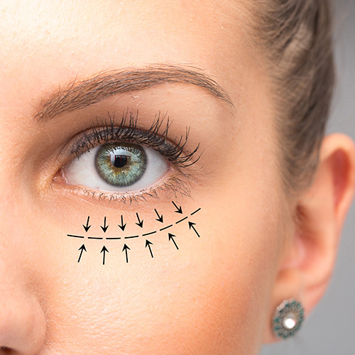
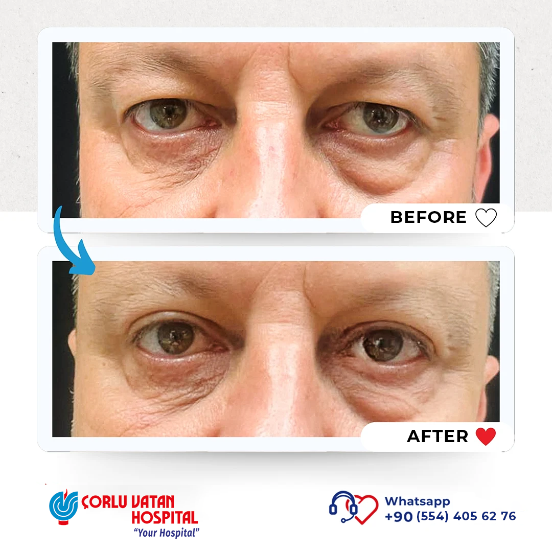
The skin around the eyes is the thinnest skin tissue in the body. For this reason, especially with age, the fat bags under the eyes become visible as the structures that hold the lower eyelid weaken. In transconjunctival lower eyelid surgery, these fat bags can be removed through a method where an incision is made from the inner side of the lower eyelid. This way, the appearance of the lower eyelid can be improved without a scar on the outer part of the eyelid. It is very important that the patient is suitable for this type of lower eyelid surgery. This surgery is not recommended for patients with a negative eye vector. After a detailed evaluation of the patient's condition, the most appropriate method for the patient is determined, and if the specific case is not suitable for this type of surgery, the method of Under-eye Lipolysis (mesotherapy with fat dissolving) is used.
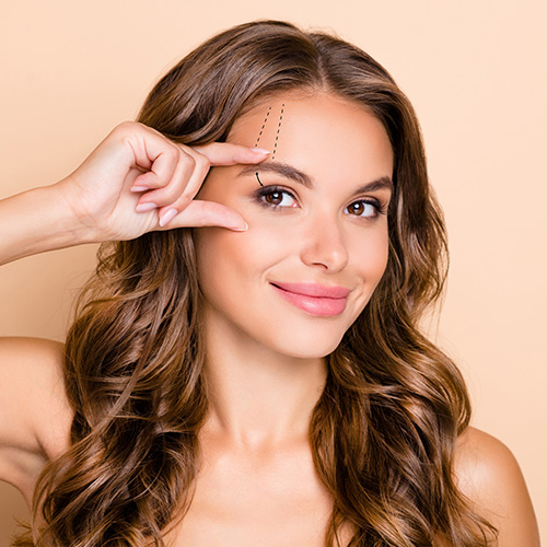
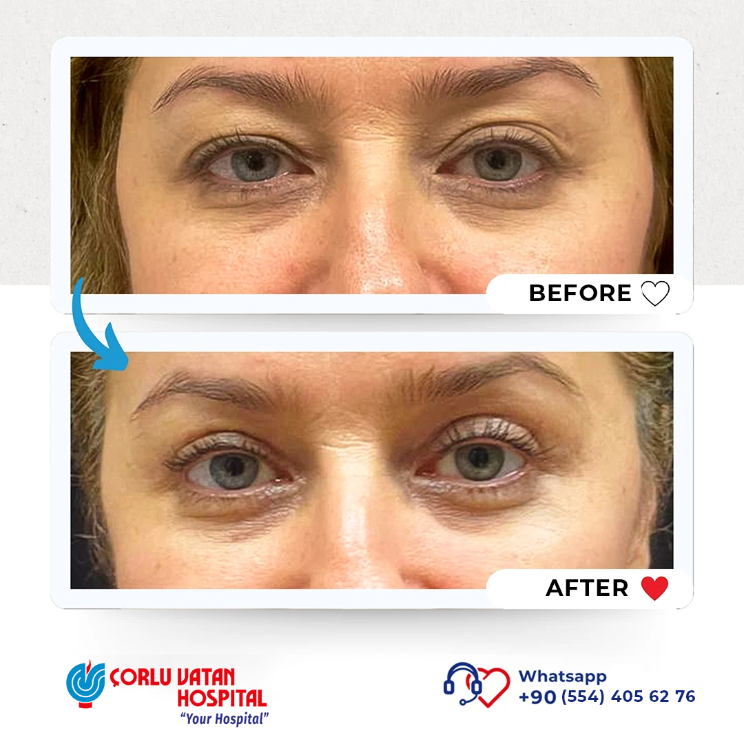
In patients where the lateral part of the eyebrow falls below the upper orbital bone, the eyebrow can be positioned higher with an incision from the inner side or just a small incision right above the eyebrow during upper eyelid surgery. A higher position of the lateral part of the eyebrow gives patients a younger and fresher appearance. In patients who do not wish to undergo surgical intervention, this part of the eyebrow can be lifted upward using absorbable meso-thread lifting methods.
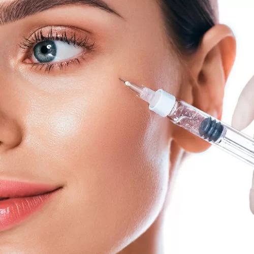
In our hospital, Botox (botulinum toxin) injection procedures are successfully performed in the forehead area and the eye area where "Crow's Feet" wrinkles are present. For patients with prominent under-eye grooves, periocular contour restoration with light filler is performed. Cheek area filling procedures are also applied to achieve lower eyelid tightening. For the treatment of dark circles and bruising in the under-eye area and to smooth wrinkles in this area, procedures such as mesotherapy are applied. For patients who have fat deposits in the under-eye area, if their case is not suitable or the patient does not wish to undergo surgical intervention, injection lipolysis procedures are successfully applied.
What Is Plasma Energy?
Plasma is known as the fourth state of matter. When sufficient energy is applied to a gas, it becomes plasma. In medicine, plasma energy is used to remove unwanted tissue or skin formations without damaging surrounding tissues.
During the procedure, energy from the device tip sublimates the superficial skin cells (keratinocytes) in the epidermis. Unlike laser or cautery, plasma energy does not spread excessive heat to nearby tissues, resulting in a lower risk of side effects.
Different energy levels are used depending on the condition:
Who Can Benefit from Plasma Energy?
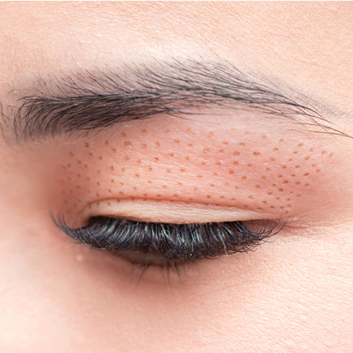
Plasma energy is a good option for patients with excess eyelid skin who do not want or do not need surgery, often referred to as non-surgical blepharoplasty. It can also improve fine wrinkles under the eyes, small skin lesions, sebaceous gland enlargements, and warts around the eyes.
In patients who previously had eyelid surgery, minor issues such as asymmetry, excess tissue, or scars can often be corrected without another operation.
How Is the Procedure Performed?
After applying local anesthetic cream, the area is disinfected and the procedure is performed in about 30–40 minutes. Small crusts form and usually fall off within about one week, revealing new pink skin.
Sun protection for about 5 weeks is essential. Mild swelling may occur for 2–3 weeks. Usually one session is sufficient, although additional sessions may be needed in some cases.
Are There Side Effects?
The main possible side effect is hyperpigmentation, which can occur without proper UV protection. With good sun protection, this risk is minimal.
How Long Do the Results Last?
The results typically last around 2–3 years.


 Phone
Phone